PHYSIO STUDY

Improving global health through universal access to physiotherapy knowledge.

December 12, 2017

physiotherapy:a boon or a curse | a boon or a curse | Is Science Boon or Bane

physiotherapy:a boon or a curse | a boon or a curse | Is Science Boon or Bane

phsiotherapy keep's you moving....🏃
Hippocrates ( Master of medicine)😎 was the first one to root the 0importance of physiotherapy 💪 in the year 460 BC, Hippocrates introduce the idea of manual manipulation for pain relief. Since then physiotherapy has evolved from simple massage to complex range of therapies.

It was in the 1950s that the physiotherapy has started outside the hospital and the field has markely increased.

It has got specialised in cardio-pulmonary💛, skin, neurological and sports therapy🚴.
Now this field has come a long way from the early nineteenth century to this date. It also include ortopedic condition such as fractute, joint disorder or dislocation, amputation, back and neck pain, arthritis and post operative management have become a more cahllening task for physiotherapist to improve the strength💪, range of motion🙌 and endurance, joint mobilization and modalities to reduce pain.
In neurological case like stroke, parkinson's disease, cerebral palsy and spinal cord injury.
A very torturing fact about being a physical therapist is that the old ancient belief on physiotherapy as a massager , this is due to lack og knowledge and awareness.

During holiday's i go back to my place my hometown, One of my neighbour aunty use come and meet me to know about my course. As soon as i start to speak " she answered me" hmm...!! in these field you have to massage full body of the patient or like the person who runs in the middle of the field when a player get injured right. Hearing those words made me speechless but i smiled And said " a physiotherapist  is the one who brings back your normal life when you suffer from movement disorder. Remember you had got stuck your neck and you went for physiotherapy treatement.

 "AFTER ALL MOVEMENT IS THE MEDICINE FOR CRAETING A CHANGE IN THE PERSON LIFE." 

December 09, 2017

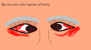
Panda sign | Panda sign in head injury

Panda sign / Raccon sign

It is called as peri orbital eccymosis.
indicate: 
draining of blood down to the periorbital soft tissue due to fracture at the base of the skull.
It can be unilateral and bilateral suggest highly severe basalar fracture.
sign:
It may accompanied by battel sign .
They may not appear after 2-3 day if the injury. 
Advice:
It is recommended that the patient should blow their nose cough vigorously or strained less to prvent further tearing of the meninges.

Panda sign,panda sign in head injury
Panda sign

December 06, 2017

Battel sign / Mastoid ecchymosis

Battle sign  0®  Mastoid ecchymosis

It is an presence of bruising  over the mastoid process.
As we know most of the extra vascular section of blood go along the path of posterior oricular artery.
Indicate:
* fracture of the base of the posterior portion of the skull
                                or
* may indicate any underline brain trauma
- This sign usually will take at least one day to appear after the initial trauma causing boggy and discoloured tissue behind ear and over the mastoid process.

   bruises is present behind the ear were the mastoid process is present.

December 05, 2017

Neurological special test | Purpose | Romberg test|special test for neurological examination

Neurological special test 
(a) Romberg test
This test is used in the examination of neurological function. This is based on the premise that person require at least two of the following three senses to maintain balance that is proprioception, vestibular function and vision.
The romberg test is used to investigate the cause of ataxia.
Procedure: 
Ask the subject to stand erect with the feet together first with eye open and then with eye closed. Make the subject to stand near to the wall or the examiner to stay close to the subject to prevent him or her from falling or hurting.
Watch the movement of the body if the subject start sway within closed eye then the test is considered as a positive sign.
Note: 
* A positive romberg test suggests that the ataxia is sensory in nature.
* If the romberg test is negative it suggests that the ataxia is cerebral in nature.

November 05, 2017

Management For Muscle Tone

Management For Muscle Tone

The management is done by different techniques and approach based on the type of lesion it can be UMN (upper motor neuron) or LMN (lower motor neuron) eg: spasticity, dystonia

muscle tone management,abnormal muscle tone management
Muscle tone .
(a) Medical management
 BOTOX
 Baclofen
 Clonidine
(b) Surgical management
 Tendon release and transfer
 Osteotomy
 Deep brain stimulation
(c) Physiotherapy management
Muscle tone was managed by rood’s approach. It is an extroceptive technique which was proposed by Margret Roods in 1970.

It was based on two techniques

1. Facilitatory techniques: Due to LMN lesion the muscle will go for flaccid, so this facilitatory technique is used to increase the muscle tone

(a) Light moving touch:-
Stimuli: light touch
Procedures: It is done with a fingertip, camel hairbrush or cotton swab and apply 2 times per sec for 10 times for 3-5 strokes and allow 30 seconds of rest between strokes to prevent over stimulation

(b) Fast brushing:-
Stimuli: brush
Procedures: Apply it over the dermatomes to stimulate the muscle which supplies the myotome for 3 to 5 sec and repeat after 30 seconds. Facial brushing is inhibited in high cervical brain stem or spinal cord lesion to prevent autonomic dysreflexia.

(c) Icing:-
Stimuli: quick icing
Procedures: Ice is applied the skin in 3 quick swipes and it is given from insertion to origin of muscle bulk.

(d) Proprioceptive Facilitatory techniques:
Heavy joint compression: I can be given with weighted cuff or sand bags. It helps to facilitate co-contraction activity of muscles

Stretch: Activates the proprioceptors in selected muscles and imply the principle of reciprocal innervations.

Tapping: This stimulus acts on the afferent of the muscle spindles and increases the tone of the underlying muscles.It can be given on the muscle bulk with the fingertips or percussed 3-5 times

Vestibular stimulation: The vestibular system is found to activate the antigravity muscles and their antagonist muscle before the stretch reflex of the muscle spindles. It can be used to obtain extensor pattern in neck, trunk and extremities

2.Inhibitory techniques: Due to UMN lesion the muscle will go for spasticity and to reduce it inhibitory technique can be used

(a) Neural warmth: Affect the temperature receptors in hypothalamus. It stimulate the parasympathetic nervous system and relieve hypertonia, spasticity, rigidity

(b) Gentle rocking / shaking: Shifting the weight forward and backward, progressing to side to side then diagonal patterns, helps to reduce the hypertonia

(c) Tendinous pressure: Manual pressure applied to the tendon insertion of a muscle; can be used in spastic or tight muscle

(d) Maintain /prolonged stretching: Positioning in the elongated position to cause lengthening of the muscle. Spindle to reset the afferents of the mm spindle to a longer position so they become less sensitive to stretch

(e) Prolong icing: Helps to reduce the spasticity of muscle to maintain normal tone

(f) Slow stroking: Patent should be on prone lying while the therapist provides a rhythmical, moving deep pressure over the dorsal distribution of the posterior rami of the spine; done from occiput to coccyx and alternated and should not exceed 3 minutes because it causes a rebound phenomenon


Muscle tone and Physiology of muscle tone | muscle tone definition,effect and physiology

Muscle tone & Physiology of muscle tone | muscle tone examination

Muscle tone is defined as an increase in the resistance of a muscle tone during passive range of motion.
                                               
                                                       OR                                                                                                                                   
Muscle resistance to passive stretch during resting state.       

Physiology of muscle tone

physiology of muscle contraction,physiology of muscle tone
physiology of muscle tone

- Tone is responsible to maintain the posture
- Muscle consist of muscle spindle and each spindle contain specialized muscle fibers they are 
intrafusal fibers and extrafusal fibers
The intrafusal fibers are (a) Nuclear bag fibers
                                       (b) Nuclear chain fibers
 In nuclear bag fibres their nuclei are clustered together in a bag like enlargement near the center of the fibres.


- In nuclear chain they donot have any enlargement and their nuclear chain has been arranged in chain fashion

- Thus both nuclear fibers have the contracting ability
- And the nuclear bag have greater diameter than the nuclear chain. So a typical muscle spindle might have 8 nuclear chain and 2 bags

- In this nuclear chain is attached to the nuclear bags, which in turns attached to the endomysium of the extafusal muscle which is the large contractile fibers.

- Thus intrafusal muscle consist two types of fibers which has both motor and sensory innervations and one or two gamma motor neuron

- So firing of gamma motor neuron will stimulate the intrafusal muscle fibers to contract
- Which activate the reflex action

- And an impulse from intrafusal muscle fibers passes through afferent fibers (1o sensory nerve fibers) and reaches the anterior gray horn of spinal cord

- Which stimulate alpha motor neuron
- Sending impulse to extrafusal fibers through efferent fibers and this will result in the contraction of muscle fibers

Effect:

Due to lesion in the UMN & LMN cause abnormal muscle tone:
(a) Hypertonia : Increase in the muscle tone on passive movement.eg: spasticity , rigidity (lead pipe and cog wheel)

(b)Hypotonia : Decrease in the muscle tone on passive movement. eg: flaccidity, floppy


(c) Myotonia : It is a congenital disease characterized by continuous contraction of muscle and slow relaxation even after the cessation of the voluntary action

October 26, 2017

Myasthenia gravis pathology | Myasthenia gravis pathogenesis

Myasthenia gravis
Myasthenia gravis is a neuromuscular disorder characterized by weakness and fatiguing of some or all muscle groups, weakness worsening on sustained exertion, or towards the end of the day, relieved by rest.

Aetiopathology
- In humans MG cause reduction of acetylcholine receptor sites in the postsynaptic folds
- Antibodies bind to the receptor sites resulting in their destruction. These antibodies are referred to as acetylcholine receptor antibodies.
- Reduced receptor synthesis and increased receptor destruction as well as well as the blocking of receptor response to acetylcholine is responsible for the disorder.
Myasthenia gravis Pathology,Myasthenia gravis defination
Myasthenia gravis
Clinical features:
Fatigue
Muscle wasting
Weakness of facial muscles
Ptosis
Nasal quality to speech
Weakness of jaw muscle – mouth remain open
Dysarthria
Dysphonia
Dysphagia
Weakness of neck muscle
Most commonly proximal muscle are affected

Sub division of clinical features:
 Class 1-          ocular muscle only
Class 2-           mild generalized weakness
 Class 3-          moderate generalized and mild to moderate ocular-bulbar weakness
Class 4-          severe generalized and ocular-bulbar 
                       weakness
Class 5-          Myasthenic crisis

Total: Class 1 (20%)
Class 2,3,4,5 (80%)

Management
Medical treatment:
 Anticholinesterase drugs 
 Steroids
 Immunosuppressants other than steroids
 Thymectomy 

Physiotherapy treatment
(a) Goals
- Myasthenia patient should find optimal balance between the physical activity and rest
- It’s not able to cure the weakness by active physical training, so low to medium intensity training is     recommended
- Enhance ability to function daily
- Decrease risk of falling
- Completion of functional tasks and maintenance of independence

(b)General exercise programs
- Should be done progressively
- Range of motion (flexibility) to light resistance to full resistance – Start with lower prescription : 3     sets of 5 reps
- The primary goal of therapy is to build the individual's strength to facilitate return to work and             activities of daily living
- Do not overdo resistive training to the point of fatigue

Type of exercise for strength are as follows :
Aerobic Exercises
Strength exercises 
Swimming
Postural exercise
Breathing exercise

(c)Postural exercise
-Important in assisting with breathing, speaking and swallowing
-Keeps bones and joints in the correct alignment so that muscles are being used properly
-Prevents fatigue because muscles are being used more efficiently, allowing the body to use less   energy

(d)Breathing exercise
- Help improve lung function
- Include inspiratory muscle training
- Pursed lip breathing
- Diaphragmatic breathing
- These exercises can improve respiratory endurance as many people with MG have affected                   respiratory muscles

(e) Exercise intensity
- The therapist has to look for the vitals after each sets of exercise
- During the time of exercise the symptoms should not be worsen
- Exercise peak dose is taking pyridostigmine

October 24, 2017

role of doctor's and physiotherapist


                                    role of doctor's and physiotherapy

                  Role of                                                                                   

-Monitoring and providing health care service to patient
-To find out the causes of the disease
-Provide medication to depress the disorder
-After injury for proper functioning of the internal system of      the   body surgeon hands play an important role 


what do physiotherapist do

         Role of

-Proper physical examination
-To prevent disability and deformity
-Educate training and transferring
-To improve tissue extensibility, increase ROM, relaxation          and  mobilize the joint the physiotherapist hand is skilled


--------------------------------------------------------------------------------------------------
So be proud of what you are and what you want to be. Your dream is your signature
-----------------------------------------------------------------------------------------------------------


October 22, 2017

ataxic gait defination | ataxic gait symptoms | ataxic gait meaning | ataxic gait causes | ataxic gait treatment

ataxic gait defination | ataxic gait symptoms | ataxic gait meaning | ataxic gait causes |ataxic gait treatment

Definition:
A loss of normal capacity to modulate fine motor movement, the patient complaint of unsteady and uncoordinated movement and lack of balance during movement it is due to cerebellar dysfunction.

The ataxic gait are of two type:

(a) Sensory gait: In ataxic gait the cerebellar are intact but the afferent fibers are     affected i.e sensory input

Sign and Symptom

-Typically the patient have lost proprioceptive and sensory input in lower limb

-They are unaware of their limb positioning

-Thus at the time of walking, they take step that are higher than necessary, while careful monitoring the ground

-Only patient with sensory ataxia slap on the floor to increase the sensory input

-And although their stance is as wide based as that of cerebellar ataxia, only sensory ataxia patients present with positive Romberg’s (swaying and falling after loss of compensatory visual input).  

(b) Cerebellar gait : This is unsteady and staggering gait towards the side of the lesion due to cerebellar lesion.

Sign and symptoms

- It is accompanied by swaying to one side or the other, so that patients often look for something to lean on – a cane, a bed rail, or even the wall.  
 -When attempting to walk tandem (heel to toe gait) it fails to maintain balance.
-Stance is also widened, but this is not enough to prevent staggering.
-Titubation while standing (fore-and-aft tremor of head and trunk) worsens considerably when patients are asked to close feet and narrow base, causing fall.
-Thus opening (or closing) the eyes neither improves nor worsens stance (negative Romberg’s)
- Hence, it differs from sensory ataxia since it is associated with other signs of cerebellar deficit, such as dysmetria , dysarthria, nystagmus, hypotonia, and intention tremor

PT management

A. provide psychological support
Give positive motivation
Gain patient confidence
Include group therapy
Explain goal and importance of exercise

B. postural correction
Use of pillows, pads to prevent abnormal posture
Use of mirror for visual feedback
Braces to correct to maintain the correct trunks

C. Sensory re-education
Use of sensory training technique like stroking, brushing and tapping
Foot splint with straps
Function stimulation

D.co-ordination and balance exercise
Non-equilibrium exercise: finger to finger exercise, finger to nose exercise, heel to shine exercise, altered pronation  /supination
Equilibrium exercise: stand with normal BOS, stand with eye open/close, stand on one leg, sit to stand, wall squatting

E. gait training
Parallel bar walking using mirror as a biofeedback
Weight shifting
Walking side way
Walking in uneven surface with support and later progress without using support

Improve heel to toe gait

October 20, 2017

waddling gait / myopathic gait / gait training / myopathic gait treatment / waddling gait treatment


waddling gait / myopathic gait
 Introduction : -
 It is a bilateral pelvic girdle weakness, typically seen in muscular dystrophy characterized by weakness of the proximal muscle of the pelvic girdle mainly the abductors of the hip.
There will be hyperextension of the trunk with shoulder thrust backward and abdomen been protuberant.

Causes

  •  Duchenne’s muscular dystrophy
  •  GB syndrome
  •  Spinal muscular atrophy
  •  OA of hip

Role of hip abductors – in waddling gait
The gluteus medius originates on the ilium (between ant. and post. gluteal lines), eventually terminating on the lateral surface of the greater trochanter.
Its contraction pulls the two insertion sites toward one another, thus elevating the opposite side of the pelvis.
 Its weakness causes contralateral sagging of the pelvis (Trendelenburg Sign)


Gait analysis
During stance phase: The weakness of proximal muscles of hip girdles, will interferes with the stability of the pelvis during walking
During swing phase: The failure to stabilize pelvis, it will produces exaggerated rotation of the pelvis with each steps
The hip are slightly flexed as a result of weakness of hip extension and there is an exaggerated lumbar lordosis.

PT Management

A. Strengthening programs

  • Active ROM to the hip joint: the movement involves hip flexion, hip extension, hip adduction, hip abduction and rotation
  • Primarily target the muscles that are responsible for gait, weakness of  muscles can lead to variety of abnormality.
  • The muscle to be strength while walking is gluteus maximus and  hamstring  for hip extension, quadriceps for knee extension, soleus &   gastrocnemius   for ankle planterflexion and dorsiflexion to step forward.
  • Progressive resisted exercise using weight cuff, theraband, resistance tube  
B. Functional balance exercise
Static exercise:      Sit to stand
                               Stand on both leg with or without support
                               Stand with eye open and close
Dynamic exercise: Straight walking
                                Tandem walking
                                Side walking      
C. Gait training

  •  Parallel bar walking by placing a mirror in front of the patient this will provide a feedback to the patient to correct the postures.
  • Make patient to walk without support
  •  During walk promote heel strike at initial contact with the floor
  • Prevent hip dropping and stabilize the pelvis

October 19, 2017

gluteus maximus gait I lurching gait


gluteus maximus gait

It is also known as lurching gait characterized by posterior leaning of the trunk at heel strike in order to keep hip extended during stance phase.


Causes

  •     Muscular atrophy
  •     Spinal muscle atrophy

Clinical features

  •     Weakness of quadriceps amd gluteus muscles
  •     Tight hamstring muscles
  •     Compensatory backward movement of trunk to maintain COG
  •     Pelvic dropping

PT Management
a. Isometric strengthening exercise of the gluteus muscles:

Ideal position: supine lying on the bed
Ask the patient to contract his buttocks and hold for 30 sec and relax.
                                         OR
The therapist hold the patient leg and passively flex hip up to 15o with hip abduction and now the patient is asked to contract the buttock.

b. Stretching of the contracted muscles like:

  •      Hamstring muscle
  •      contracted side trunk stretching
  •      Trunk ROM to maintain mobility

c. Gait training

  •     observe while walking the trunk should not lean backward
  •      Parallel bar walking by placing a mirror in front of the patient this will provide a feedback         to the patient to correct the postures. 


October 18, 2017

Choreiform Gait Physiotherapy Mx

Introduction

It is irregular, jerky, involuntary movements in both upper and lower extremities seen with certain types of basal ganglia disorder.

Clinical feature

  •         facial movement including grimaces
  •          head turning to shift eye
  •          sudden jerky movement of upper and lower extrimities
  •          unsteady gait
  •          abnormal reflex
Gait analysis

  •          it is dance like movement characterized by irregular, non repetitive or rhythmic
  •          it is wide based gait, with slow leg raising and knee flexion associated with upper limb movement
  •         this cause abnormal posture and the walking become difficult

PT management


  •         Correct the abnormal posture by positioning or by using splint: to prevent deformity
  •          Stretching of the contracted muscles: to reduce the spasticity
  •          Soft tissue and joint mobilization: to maintain the joint mobility
  •          Improve muscle tone

    Improve co-ordination and balance

a.  Non – Equilibrium exercise: - finger to nose
                                                           -finger to finger
                                                           -alternative pronation/supination
                                                           -tapping of foot
                                                           -heel to shine
         
          b. Equilibrium exercise : - standing with normal base of support
                                                 - standing with eye open / closed
                                                 - turning
                                                 - sit to stand

-         Gait and balance training

a.  Parallel bar walking using mirror as a biofeedback
b.  Weight shifting
c.   Walking side way

d.  Walking in uneven surface with support and later progress without using support


October 17, 2017

scissoring gait physiotherapy management


scissoring gait

This type of gait abnormality is associated with spastic type of cerebral palsy or upper motor neuron lesion.

Features of the following gait:


  •  it is a progressive contractures of all the spastic muscle
  •  causing rigidity and excessive adduction of the leg in swing   phase
  •  in which hip will be adducted and internally rotated
  •  knee in flexion
  •  and ankle at plantar flexion
  •  thus a complicated assisting movement of the upper limb is seen during walking
  •  inward pointed toe while walking

Gait analysis


  •  due to contracture of muscles at hip, knee and ankle it will result in excessive adduction and crossing of leg in scissor manner may touch while walking
  • subject walks on toes because of spasticity of tendoachilis
  • during walk to maintain balance they try to lift the arms and hands outwards away from the body
   PT management

To improve muscle power
 Electrical stimulation
 Strengthening exercise of weak group of muscle by resisted        exercise
Strengthening of knee extensor muscle helps to improve stride length
Aquatic therapy

To prevent contractures to reduce spasticity
To reduce the flexed postures of the body the patient should be advice to lying in prone position
Encourage to sit near the corner side of hall it will reduce the spasticity of muscle and helping the child to sit in correct position
Proper relaxation positioning
Active ROM exercise of the entire joint
Gentle stretching to increase the ROM

 Gait and balance training
First make the patient to walk with support on even surface and the progress to an uneven surface
Improve the heel to toe gait pattern
Improve side stepping
Increase the BOS during walking and progress from wide to narrow BOS
It should be noticed while the leg is kept slightly apart from each other