PHYSIO STUDY

Improving global health through universal access to physiotherapy knowledge.

Showing posts with label Orthopedic. Show all posts

September 18, 2018

Intervertebral disc prolapse physiotherapy management|pivd physiotherapy treatment

Intervertebral disc prolapsed Medical & Physiotherapy management

a) Medical management

  •  Rest             
  •  Non anti-inflammatory Antipyretic drugs

b) Reduction

Bed rest and two weeks skin traction if the pain is not reduced by corticosteroid injection

c) Chemonucleosis

Dissolution of nucleuc pulposus by percutaneous injection of proteolytic enzyme.

b) Surgical Management

Hemi laminectomy

In this one side of the whole lamina is removed

Laminectomy

In the spinous process both sides of the lamina is removed.

Laminotomy

In the lamina a hole is made for wider exposure

Fenestration

To expose the affected spinal canal, the excise the ligamentum flavum bridging the two adjacent lamina

Indication

  • No reduction in the symptoms of cauda equina
  • Persistence pain and no reduction in sciatic tension after 3 weeks
  • Neurological deterioration 

PHYSIOTHERAPY MANAGEMENT

Before planning for any function position to reduce pain should look for two bias:
Extension bias: Patient symptoms decrease in extension and provoked in flexion it shows a case of intervertebral disc prolapsed.
Flexion bias: Patient symptoms are decrease in flexion and increase in extension that shows a case of spinal stenosis, spondylolisthesis.

Acute phase

Aim

  • To relieve patient from pain
  •  To promote muscle relaxation
  •  To reduce inflammatory sign
  •  Patient education
  •  Prevention


1.Controlled rest
  • Postural correction by avoiding flexed posture
  • prevent prolong / long time sitting with any back support
  • Avoid heavy weight lifting object from the floor 
  •  Advice the patient to use local support in form of corset:Lumbosacral belt, Abdominal blinder, Tap etc
  • To prevent worsening of the symptoms, the patient is asked to take rest on a hard bed.
  •  Walking is advised to promote lumbar extension which will stimulate the fluid mechanism in the body to heal & reduce soft tissue swelling.
  •  If the patient present with inability to straighten up, make patient lie on prone position with 2-3 pillow under the abdomen. As the pain subside remove the pillow and place under the thorax, by this nucleus pulposus is shifted forwards and relieve pain and again lordosis


2.Use of modalities
  • To reduce pain and Spasm: Moist pack is used
  •  To reduce Inflammation: Cryotherapy is done
  •  To relieve pain in acute and chronic phase: TENS is used
  •  To increase the extensibility of the connective tissue: Ultrasound is used
  •  To relieve nerve compression: Mechanical traction is used


3.Initial exercise in IVDP
- For posteriolateral protrusion:
Passive extension
Lateral shift correction
- For anteriolateral protrusion:
Passive flexion
Active ROM exercise
Maintain mobility
Patient education

Sub-acute phase

  • Same exercise in acute phase
  • Cat and camel exercise: to emphasize on anterior pelvic tilt foe maintaining extension of spine
  • Back isometrics exercise


Chronic phase

a) Gentle active pain free ROM exercise


b) Stretching and flexibility exercise


back strengthening exercise

c) Core stability exercise

  •  Abdominal curls           
  •  Wall squat
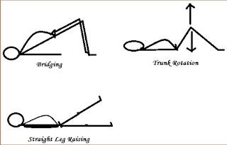
  •  Straight leg raising
  •  Plank
  •  Side plank

 d) Strengthening exercise

  •  Bridging
  •  Knee to chest
  • Trunk rotation
  •  Back isometrics


September 16, 2018

prolapsed intervertebral disc|pivd definition|pivd stages|pivd types|intervertebral disc prolapse

Intervertebral disc prolapsed/ IVDP

Definition

It is the most common cause of back pain in individuals. In IVDP there is displacement of disc material (Nucleus pulposus or annulus fibrosis) beyond the intervertebral space.
Other name used is prolapsed disc, herniated disc.

Intervertebral Disc Prolapse,Physiotherapy Management
Intervertebral Disc 


 There are four stages of Intervertebral disc prolapsed
1. Buldging
It is the early stage of disc protusion, only the disc is stretched and does not return back to the normal position when the pressure is relieved.
2. Protrusion
In this stage there is prominent disc bulge, were the nucleus pulposus has spilled out in to annulus fibrosis and barely some fibers remain inside.
3. Extrusion
In this stage nucleus pulposus has completely spilled out of annulus fibrosis and protruding out of the disc fibers.
4. Sequestration
There is complete break off of annulus fibrosis and spread into the surrounding (epidural) area.

The two types of disc lesion

Self contained disc lesion
Disc lesion with nuclear lesion
Also known as Contained disc
Also known as Non contained disc
Which means the nucleus pulposus remain contained within the annulus fibrosis.
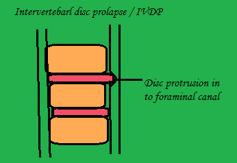
This means herniation or ruture of disc by disc protrusion into central or foraminal canal.

Common site of disc protrusion

C5 – C6  :- cervical lesion
L4 – L5 :-Lumbar lesion

Causes : -

Increasing age, sitting for long hours, poor and inadequate strength of trunk, obesity, postural stress, repetitive lifting and twisting, heavy manual labour

Clinical sign and symptoms
In early stage
a)Severe low back pain
b)Radiating pain to the buttocks and b/l lower leg
c) Pain increase during coughing and huffing
d)Lower back muscles go for muscle spasm
In later stage
a) Sensation impairment, tingling and numbness over bilateral lowerlimb
b)Muscle weakness and atrophy in later stage
c)Loss of bowl and bladder control in case of cauda equine syndrome
d)neurological symptoms: paraesthesia, numbness, muscle weakness, cauda equine syndrome
e)Deformity: Flat /  khyphotic lumbar spine, Scoliosis

Lumbar examination

Clinical examination
  • Posture :- Stand rigid, flattened lumbar spine, flat foot
  • Movement :- Unable to bend forward, muscle spasm over paraspinal area.Special examination
  • SLR
  • Lasegue
  • Femoral stretch test
  • Well leg raising test
  • Neurological test

Don't Forget to Share this Post ......

February 02, 2018

Fracture healing | fracture healing stages|stages of fracture healing

 Fracture healing


Definition
        A process of regeneration and repair ocurring across the fracture site passing through different phases of healing.

The 5 different stages of fracture healing are : 
1) Stage of haematoma formation
2) Stage of granulation tissue
3) Stage of woven bone formation
4) Stage of lamellar  bone formation
5) Stage of remodelling / consolidation

1) Stage of haematona formation
~ It is a simple stage of fracture healing as the bone fractured, bleeding occur immediately, the haematoma which is formed act as a vehical delivering the requried material for union and clearing the unwanted debris by chemotaxis.
If this stage is deficient as in open fracture healing, the fracture fails to unite.
2) Stage of granulation tissue 
~ With in 8 hours of fracture there is inflammation which subsequently leads to subperiosteal and endosteal cellular proliferation.
This cells surround the broken end of the bone and try to produce cells which differentiate and organise to provide blood vessels, fibroblast, osteoblast etc. At the same time coagulated blood get absorbed and new cappillary start infiltrating this and cellular muscle.

3) Stage of woven(soft) bone formation
~ This proliferating cell which are osteogenic and chrondiogenic they start to get incooperating into fibrogenic, under the influence of bone morphogenic protien from a transforming growth factor beta and fibroblast growth factor. Therefore forming the primary woven (soft) bone.
This bone is soft as it is not fully mineralizef it occurs at 2nd & 3rd week of fracture.
4) Stage of lamellar(hard) bone formation
~ In this stage mineralization occurs and the primary woven (soft) bone is tranformed into lamellar (hard) bone. Lamellar bone is hard abd seen as a bridge or curve at the fractured site. 
This indicate an early stage of union which occurs during 3rd & 4th week of fracture.
5) Stage of remodelling 
~ Dont considered that the remodelling take place directly after the fracture unite.
Its takes months to year after unite.
Remodelling is repeat in children and the growing bone. Also slow in adult bone and almost nill in osteoporetic bone.