PHYSIO STUDY

Improving global health through universal access to physiotherapy knowledge.

September 18, 2018

Intervertebral disc prolapse physiotherapy management|pivd physiotherapy treatment

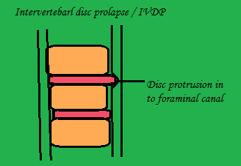
Intervertebral disc prolapsed Medical & Physiotherapy management

a) Medical management

  •  Rest             
  •  Non anti-inflammatory Antipyretic drugs

b) Reduction

Bed rest and two weeks skin traction if the pain is not reduced by corticosteroid injection

c) Chemonucleosis

Dissolution of nucleuc pulposus by percutaneous injection of proteolytic enzyme.

b) Surgical Management

Hemi laminectomy

In this one side of the whole lamina is removed

Laminectomy

In the spinous process both sides of the lamina is removed.

Laminotomy

In the lamina a hole is made for wider exposure

Fenestration

To expose the affected spinal canal, the excise the ligamentum flavum bridging the two adjacent lamina

Indication

  • No reduction in the symptoms of cauda equina
  • Persistence pain and no reduction in sciatic tension after 3 weeks
  • Neurological deterioration 

PHYSIOTHERAPY MANAGEMENT

Before planning for any function position to reduce pain should look for two bias:
Extension bias: Patient symptoms decrease in extension and provoked in flexion it shows a case of intervertebral disc prolapsed.
Flexion bias: Patient symptoms are decrease in flexion and increase in extension that shows a case of spinal stenosis, spondylolisthesis.

Acute phase

Aim

  • To relieve patient from pain
  •  To promote muscle relaxation
  •  To reduce inflammatory sign
  •  Patient education
  •  Prevention


1.Controlled rest
  • Postural correction by avoiding flexed posture
  • prevent prolong / long time sitting with any back support
  • Avoid heavy weight lifting object from the floor 
  •  Advice the patient to use local support in form of corset:Lumbosacral belt, Abdominal blinder, Tap etc
  • To prevent worsening of the symptoms, the patient is asked to take rest on a hard bed.
  •  Walking is advised to promote lumbar extension which will stimulate the fluid mechanism in the body to heal & reduce soft tissue swelling.
  •  If the patient present with inability to straighten up, make patient lie on prone position with 2-3 pillow under the abdomen. As the pain subside remove the pillow and place under the thorax, by this nucleus pulposus is shifted forwards and relieve pain and again lordosis


2.Use of modalities
  • To reduce pain and Spasm: Moist pack is used
  •  To reduce Inflammation: Cryotherapy is done
  •  To relieve pain in acute and chronic phase: TENS is used
  •  To increase the extensibility of the connective tissue: Ultrasound is used
  •  To relieve nerve compression: Mechanical traction is used


3.Initial exercise in IVDP
- For posteriolateral protrusion:
Passive extension
Lateral shift correction
- For anteriolateral protrusion:
Passive flexion
Active ROM exercise
Maintain mobility
Patient education

Sub-acute phase

  • Same exercise in acute phase
  • Cat and camel exercise: to emphasize on anterior pelvic tilt foe maintaining extension of spine
  • Back isometrics exercise


Chronic phase

a) Gentle active pain free ROM exercise


b) Stretching and flexibility exercise


back strengthening exercise

c) Core stability exercise

  •  Abdominal curls           
  •  Wall squat
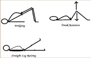
  •  Straight leg raising
  •  Plank
  •  Side plank

 d) Strengthening exercise

  •  Bridging
  •  Knee to chest
  • Trunk rotation
  •  Back isometrics


September 16, 2018

prolapsed intervertebral disc|pivd definition|pivd stages|pivd types|intervertebral disc prolapse

Intervertebral disc prolapsed/ IVDP

Definition

It is the most common cause of back pain in individuals. In IVDP there is displacement of disc material (Nucleus pulposus or annulus fibrosis) beyond the intervertebral space.
Other name used is prolapsed disc, herniated disc.

Intervertebral Disc Prolapse,Physiotherapy Management
Intervertebral Disc 


 There are four stages of Intervertebral disc prolapsed
1. Buldging
It is the early stage of disc protusion, only the disc is stretched and does not return back to the normal position when the pressure is relieved.
2. Protrusion
In this stage there is prominent disc bulge, were the nucleus pulposus has spilled out in to annulus fibrosis and barely some fibers remain inside.
3. Extrusion
In this stage nucleus pulposus has completely spilled out of annulus fibrosis and protruding out of the disc fibers.
4. Sequestration
There is complete break off of annulus fibrosis and spread into the surrounding (epidural) area.

The two types of disc lesion

Self contained disc lesion
Disc lesion with nuclear lesion
Also known as Contained disc
Also known as Non contained disc
Which means the nucleus pulposus remain contained within the annulus fibrosis.
This means herniation or ruture of disc by disc protrusion into central or foraminal canal.

Common site of disc protrusion

C5 – C6  :- cervical lesion
L4 – L5 :-Lumbar lesion

Causes : -

Increasing age, sitting for long hours, poor and inadequate strength of trunk, obesity, postural stress, repetitive lifting and twisting, heavy manual labour

Clinical sign and symptoms
In early stage
a)Severe low back pain
b)Radiating pain to the buttocks and b/l lower leg
c) Pain increase during coughing and huffing
d)Lower back muscles go for muscle spasm
In later stage
a) Sensation impairment, tingling and numbness over bilateral lowerlimb
b)Muscle weakness and atrophy in later stage
c)Loss of bowl and bladder control in case of cauda equine syndrome
d)neurological symptoms: paraesthesia, numbness, muscle weakness, cauda equine syndrome
e)Deformity: Flat /  khyphotic lumbar spine, Scoliosis

Lumbar examination

Clinical examination
  • Posture :- Stand rigid, flattened lumbar spine, flat foot
  • Movement :- Unable to bend forward, muscle spasm over paraspinal area.Special examination
  • SLR
  • Lasegue
  • Femoral stretch test
  • Well leg raising test
  • Neurological test

Don't Forget to Share this Post ......

September 13, 2018

Cardiac Physiotherapy Rehabilitation|Cardiac Rehabilitation Physio Study

Cardiac rehabilitation

Definitions
In individuals with cardiac problem it is a process of restoring the maximal level of activity for improving the functional capacity of the heart .
According to USPHS (United State Public Health Service) is defined as a rehabilitation program that involve: Medical evaluation, prescription of Exercise , education, psychological counseling.

cardiac rehabilitation physiotherapy ppt,cardiac rehabilitation physiotherapy exercises
cardiac rehabilitation
Aims of cardiac rehabilitation program
Increase functional activity & Return to safe vocation and recreation activities
Restore pt to active and productive life style & Decrease sign and symptoms such as angina
Implement safe and effective home exercise program

Rehabilitation team
Cardiologist, Pulmonologist, Physiotherapist, Occupational therapist, Dietician, Medical social worker, Psychiatrist

Indication
Most common Myocardial infarction and angina
Second commonest CABG( Coronary Artery Bypass Grafting)
And others Cardiac transplant, Pre-cutaneous angioplasty, valve replacement

Contraindication
Uncontrolled diabetes, angina, post MI infarct and arterial/ventricular arrhythmia
And other like orthostatic hypotension, recent embolism

Phases of cardiac rehabilitation
There are 4 phases of cardiac rehabilitation
A. 1st Phase [Acute phase]
It is the initial phase 0 – 7 days, the multidisciplinary team act:
1st day:-Monitoring (ECG report), Observation (position of the patient), Auscultation (Cardiac & pulmonary), Check Vitals (BP,HR,PR,RR,Temp)
2nd day:- Add exercise like upper limb exercise till 900 , chest expansion exercise like deep breathing exercise& spirometry. Along with step marching can be done.
1st-2nd day :-Patient is encourage to sit at the edge of bed or in a chair.
3rd day:-Same like above all the exercise is repeated.
2nd-3rd day :- Short distance ambulation around 6 – 9 steps is taught.
4th day :-Discontinue the BP line & check for the vitals.
5th day :- Increase the short distance ambulation around 10 – 13 steps is advised , along with stair climbing is done but before that check for the vitals.
6th day :-Six minute walk test is done and if the patient vitals and ejection fraction (50 – 60) is normal then the patient is eligible for the training of phase 2.

B.2nd Phase [convalescent phase]
After the discharge from the hospital 3 – 6 weeks of home exercise program are taught to the patient for maintaining the early mobilization & gradual increase in endurance exercise.
1st week :- Advise to have 8 – 10 hrs of rest at night and stair climbing once or twice daily.
2nd week :-Able to walk out of the doors in the gardens.
3rd week :-The daily walk can be gradually extended so by the end of 6th week the patient walk 1 – 2 miles.

C.3rd Phase [Training phase]
Both aerobic and anaerobic exercise is done.
The training that have to be followed is
1. Warm up exercise like arm swinging, trunk sideflexion, Trunk rotation with arm swing,
2. F I T T
 Frequency:- based on 6 mnts walk test
Intensity:- Moderate slowly
Type :- Walking (1RM=3 steps), jogging
Time :-20 – 30 minutes
3. Cool down period.

D.4th phase [Maintaining phase]

Exercise should be done at least twice a week for 30mnts like walking, bicycling, jogging, swimming, resisted exercise with dumbbells & pulleys.

Exercise prescription for Hypertension


September 09, 2018

Exercise prescription for Hypertension|BP

Exercise prescription for Hypertension
The competition in the world for study, job etc in individuals has caused non communicable disease i.e hypertension due to stress which was seen commonly in adults >45-50 years of age. Hypertension is an elevation in the systemic blood pressure to induce cardiovascular damage and other consequences

Exercise Goal


To achieve optimal basal physical activity and exercise status the person have to physically active and participated in other program.

1.Caution

  • controlled blood pressure
  • Regular heart rate, blood pressure, respiratory rate  monitoring
  • limited exercise

2. Aerobic exercise

  • Frequency    :2-5 times per day
  • Intensity       : -   light to moderate
  • Type              : -  rhythmic whole body exercise particularly resisted and isometrics
  • Time             : -   Minimum 15 to 30 minutes
Daily progress of the exercise is to be need based on physical examination, as higher the level of hypertension lower session of exercise iand  lower the level of hypertension longer session of exercise is done.
Progressive musculoskeletal as well as cardio-pulmonary rehabilitation has to be done.

3. Others

  • Brisk leisure activity       : -    like walking, dancing
  • Stress reducing activity  : -    Yoga, Medication, Tai chi
4. Potential pre-exercise
  • Smoking
  • Optimize nutrition
  • Fluid balance
  • Salt restriction
  • Sleep hygiene
  • Stress reduction
  • Education for regular use of medication

September 04, 2018

Clinical features in cerebral palsy|Physiotherapy management of cerebral palsy

Clinical Feature of CP

Primary : -  (due to the brain lesion)  Muscle tone (spasticity, dystonia)
                                                   Balance
                                                         Strength
                                                         Selectivity
                                                         Sensation
Secondary  :  - due to the primary impairments causing the movement disorder)
Contractures (equinus, adduction)
Deformities (scoliosis)
Tertiary : -  Adaptive mechanisms (knee hyperextension in stance)

Problems / deformities commonly seen in CP

Upper limb
Shoulder - flexion, adduction and internalrotation deformity
Elbow – flexion and pronation of forearm                                         
Wrist – flexion and ulnar deviation

Lower limb
Windswept deformity (hip joint)
Occiput side – limb in flexion, abuction and external rotation
Face side – limb in flexion, abduction and internal rotation
Knee joint
Flexion deformity
Valgus
Genu recurvatum
Ankle joint
Equinus
Valgus/ Varum deformity
Clinching of toes

MANAGEMENT OF CP

PHYSIOTHERAPY ROLE

P.T. especially when started early in life, is helpful in promoting normal motor development, and preventing deformity and contractures.

A) Medical MX
Management of Spasticity
Drugs
a. Baclofen
b. Diazepam
c. Botulinum toxin

Surgical

Tendon lengthening and transfer and arthrodesis are some of the procedures commonly performed.
Dorsal rhizotomy: which involves selective resection of posterior nerve roots from L2 to S2. It may be helpful in children with severe lower limb spasticity, with sufficient trunk control and some form of forward locomotion.

B)PhysiotherapyMX

1.Handling and positioning
2. Therapeutic exercise
- Gentle range of motion exercises
- Strengthening exercises
- Functional progressive resistance exercises
- Stretching
3.Therapeutic handling
- To influence the quality of motor response
- To use sensory information and adapt movements
4. Facilitation
- Process of intervention which uses postural tone in a goal directed activity
5. Proprioceptive neuromuscular facilitation[PNF]
PNF uses the body’s proprioceptive system to facilitate or inhibit muscle contraction. 
CP is the major the problem seen in young child, so different techniques has been and one of them is Bobath technique, roods approaches and play therapy.
Bobath technique is used to improve neuromotor development.
Roods approaches for muscle tone management by inhibitory technique (Slow rolling, prolong stretch, slow stroking) and facilitatory technique(fast stroking, quick icing, joint compression/weight bearing, stretching).
Play therapy will encourage the child to move on.
As the aim of physiotherapist should be on functional activity to make the child practice it in a real life situation.

6. Assistive and adaptive devices
Angled spoons: two handled cups
Old stools and boxes: to provide support during sitting
Standing frames: are used in the stage of mobilization
Parallel bars: for gait training
Splints, Casts and Calipers: Specially designed shoes, ankle-foot orthoses (AFO) and calipers


September 03, 2018

cerebral palsy definition | cerebral palsy types

 CEREBRAL PALSY DEFINATION / TYPES OF CP ( LITTLE’S DISEASE )

Defination
It’s
1) Persistent but not unchanging disorder of movement, tone and posture
2) due to non-progressive defect / lesion of immature brain at fetal life, infancy and childhood.
Also associated with spectrum of developmental disability such as
Ø Mental retardation
Ø  Epilepsy
Ø  Visual, hearing and speech defects
Ø  Strabismus
Ø  Cognitive dysfunction
Ø  Sensory, emotional and behavioral problems
                                                         OR                                                                     
Umbrella term covering non-progressive but often changing motor impairment syndrome that may or not involve sensory deficits that are caused by a non-progressive defect , lesion or anomaly of the developing bran and that can be in part a developmental diagnosis
First described by William Little in 1862.Then it was known as Little disease.

Classification

TOPOGRAPHIC

PHYSIOLOGICAL
1
MONOPLEGIA
1
SPASTIC
2
HEMIPLEGIA
2
EXTRAPYRAMIDAL
3
DIPLEGIA
3
ATAXIC
4
QUADRIPLEGIA
4
MIXED
5
DOUBLE HEMIPLEGIA
5
ATONIC
6
TRIPLEGIA
6
ATHETOID

TOPOGRAPHIC CLASSIFICATION
 
cerebral palsy definition ,cerebral palsy types
Topographic Classification of CP.
PHYSIOLOGICAL CLASSIFICATION
1) SPASTIC CP
Spasticity is defined as an velocity dependent increase in the physiological resistance of muscle to passive motion.
Result from damage to motor areas of the cerebrum; characterized by increased muscle tone, primarily of flexors and internal rotators, which might lead to permanent contractures and bone deformities.

SPASTIC DIPLEGIC
Involvement of legs more than arms often associated with premature birth. Only 11-20% are severely impaired.MR not so profound.

SPASTIC QUADRIPLEGIA
Involvement all four limbs, arms at least severely affected as leg. Severely impaired and MR. Often have bulbar symptomatology.

SPASTIC HEMIPLEGIA
Involvement of arm and leg on one side(arm > leg). Motor handicaps at least likely to be disabling. Intelligences is normal to dull.

2) ATAXIC CP
Ataxia is loss of balance, coordination, and fine motor control. Ataxic children cannot coordinate their movements. They are hypotonic during the first 2 years of life. Muscle tone becomes normal and ataxia becomes apparent toward the age of  2 to 3 years. Children who can walk have a wide-based gait and a mild intention tremor .Fine motor control is poor. Ataxia is associated when there is damage to the cerebellum (centre of balance and co-ordination).

3) ATHETOSIS CP
A condition that occurs when there is damage to the basal ganglia (masses of gray matter composed of neurons located deep within the cerebral hemispheres of the brain) results in an overflow of motor impulses to the muscles. Some characteristics of this type of CP include slow, writhing movements that are uncoordinated and involuntarily.

4) MIXED CP
Children with a mixed type of CP commonly have mild spasticity, dystonia. Ataxia may be a component of the motor dysfunction in patients in this group. Ataxia and spasticity often occur together. Spastic ataxic diplegia is a common mixed type that often is associated with hydrocephalus.

AREA AFFECTED IN CP
Site of brain injury
Pathological
Cortical
Periventricular lucomalacia   » Spastic diplegic
Sub-cortical
Stroke in utero  » Hemiplegic
Periventricular
Multifocal encephalomalacia  » Quadriplegia
Basal ganglia
Cerebellar  » ataxia
Cerebellum
Basal ganglia, thalamus, putamen  » dyskinetic
Brain stem