
INTRODUCTION ;
Bony and cartilaginous framework of the body consititutes the skeleton.
Anterior view
HOMOLOGOUS PART OF THE LIMB
Upper Limb it include
 1.Shoulder girdle
2.Shoulder Joint
3.Arm With Humerus
4.Elbow Joint
5.Forearm Radius and Ulna
6.Wrist Joint
7.Hand With
(a) Carpus
(b) Metacarpus
(c) 5 Digits

Lower Limb
1.Hip Girdle
2.Hip Joint
3.Thigh With Femer
4.Knee Joint
5.Leg With Tibia and Fibula
6.Ankle Joint
7.Foot With
(a) Tarsus
(b) Metatarsus
(c) 5 Digits
The Skeleton divided in to two :
ENDOSKELETON : - It is located internally in body.in human anatomy the term skeleton usually means endoskeleton
EXOSKELETON : - In human beings the exoskeleton is very rudimentary being represented by nails & enamel of teeth only.
NUMBER OF BONES : 206
Upper Limb-----------------64
Lower Limb-----------------62
Vertebrae--------------------33
Skull--------------------------29
(Facial Bones - 14, Cranial - 14, hyoid bone - 1)
Ribs---------------------------24
Sternum------------------------1

Bony and cartilaginous framework of the body consititutes the skeleton.
Anterior view
HOMOLOGOUS PART OF THE LIMB
Upper Limb it include
 1.Shoulder girdle
2.Shoulder Joint
3.Arm With Humerus
4.Elbow Joint
5.Forearm Radius and Ulna
6.Wrist Joint
7.Hand With
(a) Carpus
(b) Metacarpus
(c) 5 Digits
Posterior View

Lower Limb
1.Hip Girdle
2.Hip Joint
3.Thigh With Femer
4.Knee Joint
5.Leg With Tibia and Fibula
6.Ankle Joint
7.Foot With
(a) Tarsus
(b) Metatarsus
(c) 5 Digits
The Skeleton divided in to two :
ENDOSKELETON : - It is located internally in body.in human anatomy the term skeleton usually means endoskeleton
EXOSKELETON : - In human beings the exoskeleton is very rudimentary being represented by nails & enamel of teeth only.
NUMBER OF BONES : 206
Upper Limb-----------------64
Lower Limb-----------------62
Vertebrae--------------------33
Skull--------------------------29
(Facial Bones - 14, Cranial - 14, hyoid bone - 1)
Ribs---------------------------24
Sternum------------------------1
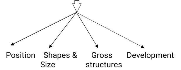
CLASSIFICATION
OF BONES : BASED ON
A.ACCORDING
TO POSITION
(1)
Axial : Bones
Forming the axis of the body.
Eg.
Skull, Ribs, Sturnum, Vertirae
(2)
Appendicular : Bones forming the skeleton of the limbs
B.
ACCORDING TO SHAPE AND SIZE
(1)
Long bone : present in
upper limb and lower limb
(2)
Short long bone : They are miniature in size,
eg.
Metacarpal & Metarsal and Phalanges
(3)
Short bone : they are small and cuboidal in shape.
eg. Carpal & Tarsal bone.
(4)
Flat bone : They are expanded & protect
the vital struct.
eg. Scapula,Sternum, Ribs, Pareital & Frontal bones.
eg. Scapula,Sternum, Ribs, Pareital & Frontal bones.
(5)
Irregular Bone : Irregular in out line.
eg. this
include Vertebrae, some skull bones.
(6) Pnumatic bone : Flat
or irregular bone possessing a hollow space within there body which contains air, eg. Ethmoide, Maxilla, Mastoid part of temporal
bone.
(7) Sesamoid Bone : it means seed like, eg: Patella,
Pisiform Bone,
(8) Warmian Bones : Sutural bones. These are found along the sutures,
Can be
mistaken for fractures.
C. ACCORDING TO GROSS STRUCTURE :-
(1) Compact (lamellar) bone - Outer cortical part of long bone which is hard.
(2) Spongy (cancellous) bone - The part of bone which is less hard.
(3) Diploic bone - Consist of inner & outer tables of compact bone.with an intervening porous layer which is occupied by a spongy substance consisting of bone marrow and diploic veins.
D. ACCORDING TO DEVELOPMENT : -
(1) Membranous (Ectochondral) bones - Which develop in membrane.


Reviewing the fundamental anatomy of the human skeleton is a great refresher, but translating the physical architecture of bone loss into a highly detailed, clinical care plan is a massive hurdle. I got completely overwhelmed last week trying to synthesize bone density markers, pharmacological rationales, and progressive mobility interventions for a complex musculoskeletal case study.
ReplyDeleteI ended up reaching out for Osteoporosis Assignment Help by Nursing Expert Writers to get my clinical reasoning properly structured. The team did an amazing job helping me align my nursing interventions with evidence-based patient safety metrics, which was a total lifesaver for getting my report finished accurately.
Every HR manager should read articles like this.best hr software
ReplyDeleteThis breakdown of the skeleton's structural classification brings me back to anatomy lectures, especially the note on how Wormian bones along the skull sutures can easily be mistaken for fractures on imaging. Keeping all these anatomical distinctions straight while balancing clinical placements is brutal, and last semester when I was juggling practical exams and a massive research paper on biomechanics, getting New Assignment Help with Urgent Assignment Help in Sydney was an absolute lifesaver for keeping my study schedule on track. It’s always handy having a concise overview like this to quickly double-check bone categories and basic structural counts during quick revisions.
ReplyDeleteThis breakdown is a really handy refresher! It’s interesting to be reminded that human fingernails and tooth enamel are actually classified as rudimentary components of the exoskeleton. Going through these foundational concepts while managing clinical coursework can be quite intense, especially when balancing practical work alongside theory. Having some support through Diploma Assignment Help Australia definitely makes managing that workload a whole lot easier for students during busy semesters. It makes me wonder how much the overall balance between endoskeleton and exoskeleton functions varies across other species when you look deeper into comparative anatomy. Thanks for sharing such a clear and well-structured overview.
ReplyDeleteThis is a handy summary of the human skeletal system. I had forgotten that we have 206 bones in total, and seeing the breakdown between the axial and appendicular systems puts things into perspective. It is fascinating how bones are classified based on shape, like short bones in the carpal region versus long bones in our limbs. From a student's perspective, the clear structure and key Language Features used throughout the post make the anatomical terminology so much easier to digest compared to dense textbooks. I wonder how much the density of spongy cancellous bone changes as we age compared to compact bone. Thanks for sharing such a neat overview!
ReplyDelete